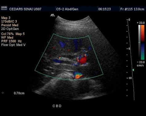
What is Abdominal Ultrasound?
Abdominal ultrasound is an anatomy procedure of a kind. It is used to analyze muscles, including the liver, gallbladder, spleen, pancreas, and kidneys, in the belly. It is also possible to analyze the blood vessels leading to each of these organs, such as the inferior vena cava and aorta, using ultrasound.
Why is an ultrasound of the abdomen performed?
To check the main organs in the abdominal cavity, abdominal ultrasounds are used. The gallbladder, kidneys, liver, pancreas, and spleen are among these tissues. If your doctor believes that you have some of these other disorders, you might undergo an abdominal ultrasound in the immediate future:
1. Clot of blood
2. Expanded organ (such as the liver, spleen, or kidneys)
3. Abdominal cavity blood
Preparations
Ask the doctor if you can continue to take your medicine and drink water before an ultrasound. Your doctor would typically advise you to have it 8 to 12 hours before because the sound waves can be blocked by undigested food in the stomach and urine in the bladder.
Risks
There are no complications from the abdominal ultrasound. Ultrasounds do not use radiation, unlike X-rays or CT scans, which is why doctors tend to use them in pregnant women to monitor for growing infants.
Results
Your ultrasound pictures are analyzed by a radiologist. At a follow-up visit, the doctor will review the conclusions with you. Your doctor can order a further follow-up scan or other examination and set up an appointment to check for any problems that have been discovered.
Consult
Dr. Manju Whig Singh provides abdominal ultrasound in greater Noida, talk to her if you have any concerns.
Services
- Level 1 or NT/NB Scans, Level 2 Scans
- Transvaginal Ultrasound
- Baby Brain Ultrasound
- Neck, Eye, Joint Ultrasound
- 3D 4D Ultrasound
- Breast Ultrasound
- MammoGraphy
- Color Doppler
- KUB
- Follicular Monitoring
- ADD TVS Ultrasound
- Sonosalpingography
- ElastoGraphy
- Pelvis Ultrasound
- Growth Scans
- Foetal Echo Cardiography
- Obstetrics or Foetal Scans
- Ultrasound Guided Procedures


