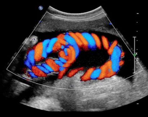
A Color Doppler Ultrasound is a scan to see how the lungs and veins carry blood. The research blends conventional ultrasound with ultrasonography from Doppler. To produce images, standard ultrasound uses sound waves that bounce off blood vessels. Doppler aims at how sound waves, like blood, are reflected away from moving objects.
Why it's done?
1. In almost every part of the body, blood clots and blocked or shortened blood vessels are found, particularly in the throat, arms, and legs.
2. Test irregular veins that cause or cause other complications for varicose veins.
3. Determine the volume of blood supply to a kidney or liver that has been transplanted.
4. Track blood supply after blood vessel surgery
What is the Procedure of Color Doppler Ultrasound?
You’re going to be sitting on a bench, usually on your stomach. A cream may be applied to the area to be examined by the doctor or technician. This helps you move across the sound waves and allows you improved performance. Then, a delicate device will be pressed against your skin. It sends sound waves through the body as they drive the device around.
Results
Your doctor will let you know the significance of all the photos. They will tell you what the photos reveal in your blood pressure and tell you the next actions to follow if you get the scan done to search for DVT.
Consult
Dr. Manju Whig Singh provides the service of color Doppler ultrasoundin 3D 4D Ultrasound, Greater Noida, and treats patients according to results.