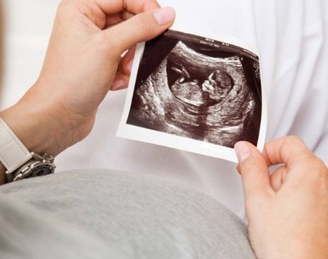
When the mother is between 23 and 40 weeks pregnant, the growth scan also called the health scan or positioning scan takes place. This examines how well the baby is developing and the uterus location.
What is the need for a growth scan?
The following are common explanations for having a fetal growth scan during pregnancy:
1. Your abdomen is smaller or bigger than expected.
2. Previous risks from conception.
3. Maternal diabetes or hypertension.
What is the procedure for a Growth scan?
Various measurements of the embryo are made during the fetal growth scan. For a growth scan, the major fetal measurements taken include:
1. Across the head, biparietal diameter (BPD) tests
2. Head Diameter (HC)-Action on the head
3. Abdominal Diameter (AC)-abdomen measurements
4. Femur Length (FL)-measures the thigh bone size
The head, abdominal, and leg measures enable the doctor to determine the weight of the fetus. Both scales are plotted against the standard range and to determine the development of the fetus on a map. Since babies develop from week to week at various speeds, a series of scans may be more beneficial than just one.
Risks
It has no ill effect on baby or pregnant women.
Results
The findings of the ultrasound will reveal whether your baby is progressing normally or suffering from reduced fetal development (FGR). Restriction of fetal growth (FGR) is a term that describes a fetus that does not develop properly before birth. It is also called the restriction of intrauterine development.
Consult
For growth scans, you can believe Dr. Manju Whig Singh. She provides you with all the checks that you need to rule out any irregularities and allows you to learn how to treat pregnancy stress.